
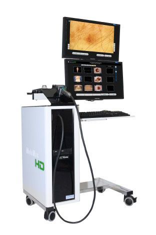
MoleMax HD PRO
High Definition Mole Mapping System
The Melanoma changes its appearance and early changes can only be detected through comparison of at least two images taken 3 to 6 months apart.A major technological advancement in Melanoma detection, the MoleMax/DermDOC system, available now at Beach Road Surgery & Skin Clinic will help alleviate your concerns.
Dermoscopic images of suspicious pigmented skin lesions can be magnified up to 100 times and then stored in the MoleMax/DermDOC data base.
At successive check-ups current images are compared to the previously stored one. This technology enables us to detect subtle changes in moles and new moles can be identified over time leading to early detection of melanoma and therefore a better chance of cure.
System advantages
- The MoleMax system was the first digital dermoscopy system that was developed in Austria in 1977.
- This system enables your doctor to examine your skin beyond its surface layers and detect changes in the skin not visible to the human eye.
- This makes diagnosis much easier and enhances the chances of detecting a malignant melanoma early.
- This quick and non-invasive examination is not only painless but also allows patients to observe examinations on the TV screen. No oil is required so a clean and comfortable examination is performed.
Total Body Photography (TBP)
Total Body Photography (also known as TBP or Full-Body Imaging) is the most effective technique for imaging all lesions on a patient’s body. Total body photography involves photographs taken of your entire skin (wearing only undergarments), allowing a complete photographic record of the location, size and appearance of your moles (melanocytic naevi). Full-body imaging is the most reliable means of accurately tracking changes over time and producing potentially lifesaving results as the images allow easy side-by-side comparison at follow-up appointments for patients in certain risk groups. With sophisticated software and superior expertise working together for exceptional accuracy, we can reveal the subtlest skin changes and detect skin cancer at its earliest stage when it is most manageable and requires the least invasive treatment.
Early diagnosis of melanoma
The risks involved with melanoma can be dramatically reduced if the disease is diagnosed early, with photography and digital monitoring, often described as ‘mole mapping’, playing a significant role in cancer prevention. This is especially the case for patients with numerous moles that require ongoing observation.
Early detection of change in moles and lesions is a key component in diagnosis and cure of melanoma, the most aggressive form of skin cancer. Advances in medical science and technology have resulted in the development of imaging systems capable of digitally monitoring moles and lesions as they change over time. Skin Cancer doctors can analyse dermatoscopy pictures, compare images, and even use computer-aided diagnosis for confirmation when regular moles are changing into potential melanoma.
Reducing biopsies
In their quest to ensure melanoma doesn’t occur, skin cancer doctors in the past have tended to remove benign moles as a precautionary measure. In fact, for every malignant melanoma removed, hundreds of benign lesions are also sacrificed. Biopsies remain an essential procedure for evaluating the condition of skin lesions, although time and resources are undoubtedly wasted when procedures are inadequate for the task at hand. At Beach Road Surgery & Skin Clinic, digital monitoring is a dermatological game-changer.
Repeat photographs are taken at either 3 or 6 months, depending on the type of lesion, and as recommended by your skin cancer doctor. Research shows that 99% of lesions that do not change over a three-month period are benign, while 96% of melanomas will demonstrate subtle changes within 3 months. Lesions which undergo change may then be recommended for surgical biopsy.
Digital monitoring for increased patient satisfaction
Patients enduring skin problems and skin diseases are understandably distressed and looking for answers. Many dermatologists believe total body photography reduces patient stress and anxiety by promoting a positive involvement in the healing process. The data-based approach means patients have information on hand when needed, allowing them to become more involved in their own care. In any case, it’s usually the patient or close family member who first notices the melanoma, and photographic data is a useful way to confirm any changes in moles or lesions, even while at home.
Digital monitoring is a relatively new procedure and now it is available to you in Eurobodalla. It’s worth noting, however, that ‘change’ is the major determining factor used when assessing moles and lesions. The sooner any change is noticed, the sooner it can be monitored, mapped or removed if necessary.
Who would benefit from Total Body Photography?
Someone who:
- has a personal or family history of melanoma
- has a personal or family history of multiple dysplastic (abnormal) moles
- has multiple (> 50) moles
- is developing new moles
- has a history of multiple non melanotic skin cancers is taking immunosuppressant therapy for various conditions


Beach Road
Surgery & Skin Clinic
Address:
116 Beach Road,
Batemans Bay, NSW 2536
Phone:
Opening Hours:
Monday - Friday: 9:00am - 4:30pm